

Our Product
Cardiac Insight, Quantified
Introducing Vocelo™ 3D Myocardial Shear Wave Elastography (3D MSWE) — exploring new frontiers in cardiac biomechanical properties. Our goal is to develop a new approach in heart failure management using our unique technology based on shear wave elastography (SWE) and ultra-fast ultrasound imaging to assess cardiac stiffness.
![]()

Our Technology
The eMyosound System: Pushing the Boundaries of Cardiac Assessment
We are developing an ultrasound-based system that uses a non-invasive approach to assessing and quantifying cardiac stiffness. Our research leverages cutting-edge 3D ultra-fast shear wave elastography and signal processing to quantify cardiac tissue elasticity, aiming to provide unique insights into the mechanical properties and function of the heart. Our innovation relies on highly integrated technology, allowing us to reliably estimate the 3D biomechanical properties of the myocardium at controlled intervals of the cardiac cycle.
Proprietary Smart Ultrasound Technology Based on our Innovative Transducer
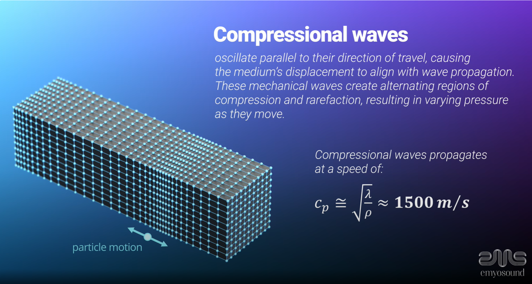
Our development of advanced ultrasound technology relies on the principles of shear wave imaging. This approach aims to assess the mechanical properties of soft tissues non-invasively, by analyzing the propagation of shear waves remotely induced by acoustic radiation force.
Our ultrasound transducer, composed of an optimized number of piezoelectric elements, is the only of its kind able to both generate and track the three-dimensional propagation of shear waves in complex biomechanical tissues such as the fibrous architecture of the heart. Our research focuses on applying this technology to achieve non-invasive and real-time quantification of anisotropic myocardial stiffness in human patients.
Disclaimer: This technology is currently in the preclinical development phase. The results presented are preliminary. Regulatory approval has not yet been obtained.
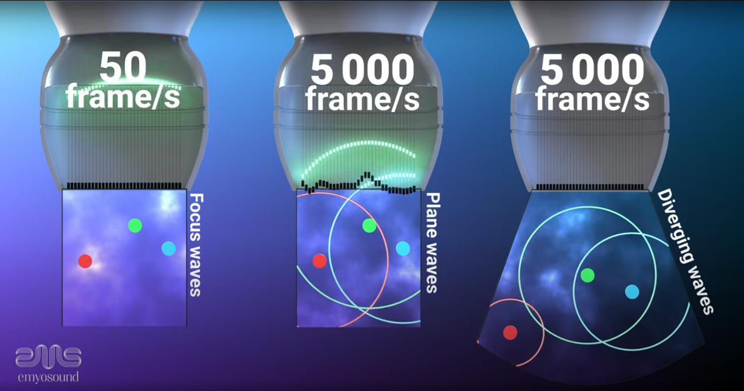
Understanding Shear Wave Elastography (SWE): How We Measure Tissue Stiffness
Shear waves — a type of mechanical wave that propagates sideways — are generated within the tissue. The speed at which these shear waves travel is directly related to the local stiffness of the tissue. Stiffer tissues cause shear waves to travel faster, while softer tissues cause them to travel slower. By measuring the speed of these waves, SWE can provide a quantitative assessment of tissue elasticity. Ultrasound transducers are used to generate these shear waves, often using acoustic radiation force. A transducer is used to track the propagation of these waves through the tissue at ultra-fast frame rates. Sophisticated software processes the data to create images or numerical values that represent the tissue’s stiffness.
3D Myocardial Shear Wave Elastography

Vocelo™: A Unique Mode to Investigate Cardiac Tissue Properties
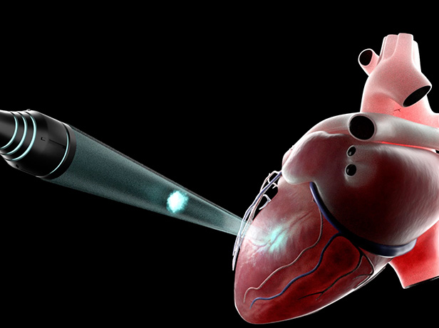
Vocelo™ is designed to direct a precise acoustic radiation force, allowing for the generation of shear waves in specific regions of interest, including the right ventricular wall, septum wall, and apex. This targeted approach aims to provide highly localized insights into myocardial stiffness.
The eMyosound system is being developed to potentially deliver real-time feedback to healthcare professionals. This feedback is intended to support the investigation of how cardiac tissue responds to different conditions, with the goal of improving the monitoring and management of heart health.
Our goal is to enhance the patient journey by enabling more personalized assessment and paving the way for future innovations in cardiac imaging. While still undergoing research and evaluation, the eMyosound system represents a novel approach with the potential to help advance cardiac diagnostics and treatment pathways.
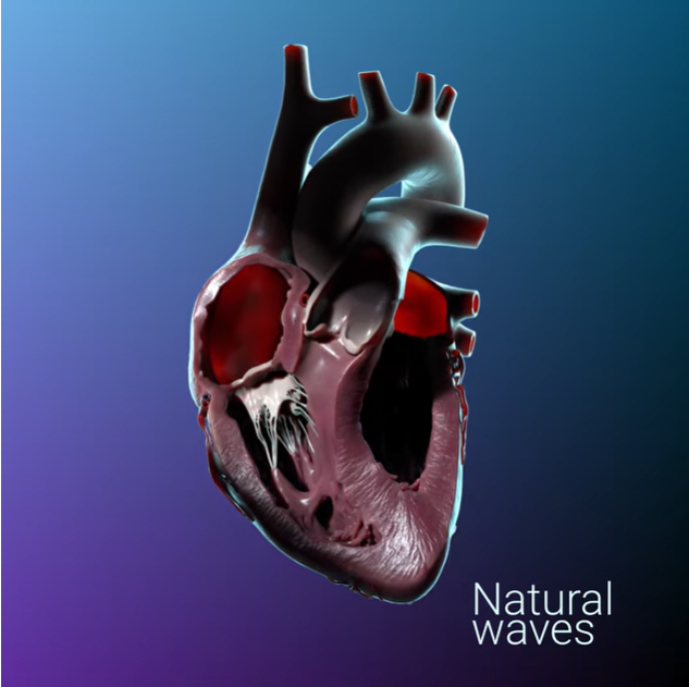
Beyond these induced waves, our innovative approach also enables the detection and analysis of the heart’s intrinsic natural shear waves. These natural waves, generated by the closing of the mitral and aortic valves, carry crucial information about the heart’s underlying mechanical properties and stiffness in a physiological state. Measuring both induced and natural shear waves provides a more comprehensive understanding of myocardial mechanics and the heart’s rigidity, offering deeper insights into cardiac function.


Let’s Connect
Whether you’re a clinician, researcher, or industry leader, we invite you to join eMyosound’s mission in transforming cardiac healthcare for patients.